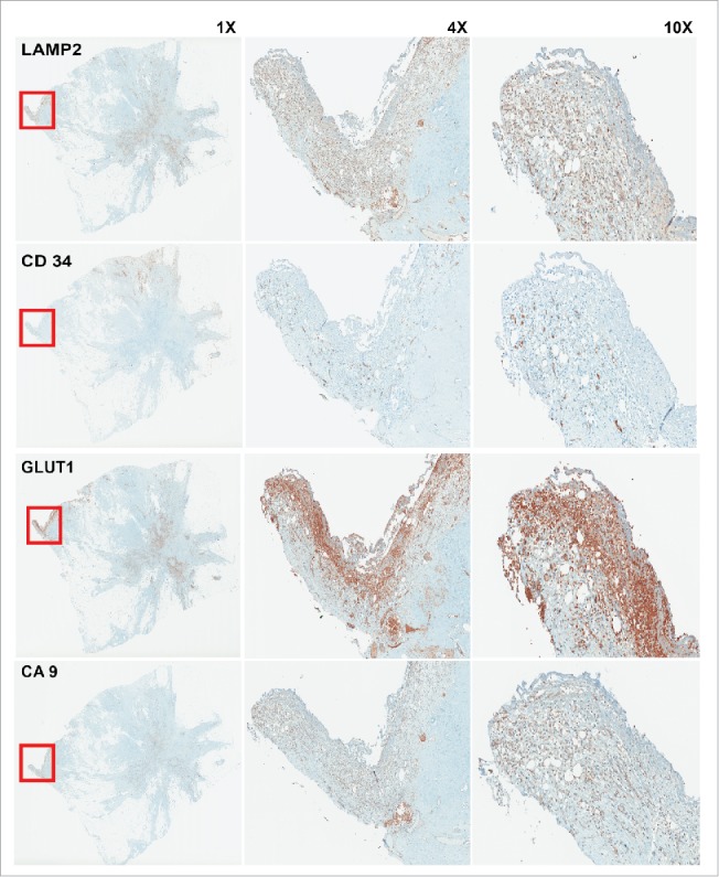
Figure 3.

IHC staining of sequential cuts of patient sample. The red box shows the area with less vasculature marked by low CD34 staining. This area is hypoxic and acidic as depicted by GLUT1 and CA9 staining. As we see this area has highest expression of LAMP2 that is a marker for acid adapted cells. Acid adaptation plays a major role in acquiring new phenotype that make the adapted cells as pioneer cancer cells that can protect themselves and other local cancer cells and also lead the invasion and migration toward adjacent tissues.
