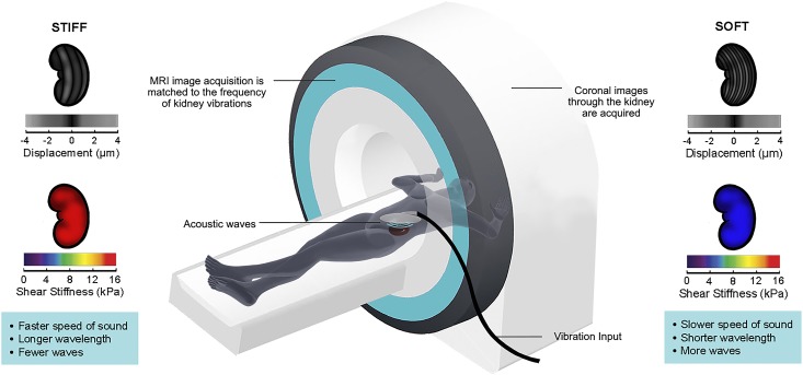
Figure 1.
Magnetic resonance elastography can be used to image transplant kidney stiffness. In magnetic resonance elastography, a gentle vibrational wave is applied on the skin overlying the kidney using an acoustic generator. The magnetic resonance imaging (MRI) scanner then images the small displacements produced by the resulting shear waves generated in the kidney. Because the waves are longer and travel more rapidly through stiff tissue, kidney stiffness can be calculated on the basis of the measured displacements and the amount of vibrational force applied. Postimaging quantitative reconstruction enables the generation of stiffness maps (elastograms) of the imaged kidney. Displacement images are shown to the left and right of the scanner. Below each displacement image is the corresponding pseudocolorized elastogram (stiffness map); blue depicts softer tissue, and red depicts stiffer tissue.

