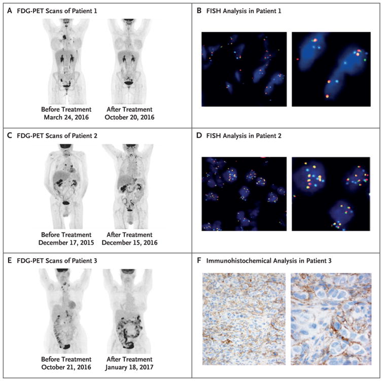
Figure 1. 18F-Fluorodeoxyglucose (FDG) Positron-Emission Tomographic (PET) Response, 9p24.1 (PD-L1/PD-L2) Fluorescence In Situ Hybridization (FISH) Analysis, and PD-L1 Immunohistochemical Analysis.
Panels A, C, and E show maximum-intensity projection images from FDG-PET scans of three patients with refractory mediastinal gray-zone lymphoma who were treated with programmed death 1 (PD-1) blockade. As shown in Panel A, Patient 1 had a complete metabolic response after treatment with pembrolizumab, with no residual hypermetabolic uptake on post-treatment imaging. As shown in Panel C, Patient 2 had a complete metabolic response after treatment with pembrolizumab, with residual uptake in the neck and upper chest that was consistent with normal-variant brown fat. As shown in Panel E, Patient 3 had a complete metabolic response after treatment with nivolumab, with diffuse uptake in the bowel that was compatible with inflammation. Amplification and rearrangement of the PD-L1/PD-L2 locus was investigated by FISH with the use of a PD-L1/PD-L2 Break Apart probe (labeled with Spectrum Red and Spectrum Green) and a CEP 9 probe (labeled with Spectrum Aqua). Panel B shows rearrangement of the PD-L1/PD-L2 locus in Patient 1, with a split green signal suggesting the presence of an inversion within this region. Panel D shows amplification of the PD-L1/PD-L2 locus in Patient 2, with evidence of multiple fusion signals (69% of cells with ≥6 copies per cell, 20% with 3 to 5 copies, and 11% with ≤2 copies). In Panel F, immunohistochemical analysis in Patient 3 shows focal membranous expression of PD-L1 in the lymphoma cells, with additional expression seen on the small background cells surrounding the malignant lymphoma cells.

