Figure 4.
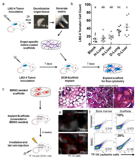
Modelling organotropism using ECM- or BMSC-functionalized scaffolds. (a) Decellularized lung and liver matrix from healthy and diseased mice inoculated with tdTomato-tagged LM-2 lung/liver targeting breast tumour cells was used to coat PCL scaffolds, and scaffolds were implanted subcutaneously in tumour-inoculated mice to detect differences in tumour cell colonization as a function of matrix coatings. Mouse image drawn by Katie Aguado and reproduced with permission from Nature Publishing Group35. (b) Matrix-coated scaffolds from diseased lungs and livers recruited more cells relative to blank and healthy coating controls as assessed by flow cytometry. Groups with different letters are significantly different (P < 0.05). Figures reproduced with permission from Elsevier77. (c) Delivery of multipotent BMSCs (CD44+, CD106+, CD14−, CD34−, CD45−, CD73+, and CD105+) on scaffolds recruit TF-1A leukemia cells to an implant site. (d) Images of H&E stained tissue sections of subcutaneously implanted 3D microfabricated polyacrylamide scaffolds (unseeded vs. BMSC seeded, scale bars = 250 μm). (e) Homing of intravenously transplanted human TF-1A cells to unseeded vs. BMSC seeded scaffolds. Confocal images of scaffolds show significantly more stained TF-1A cells arriving to BMSC-seeded scaffolds 6 hours after injection (scale bars = 250 μm). (f) Flow cytometric analysis of labeled TF-1A cells at the bone marrow vs. implanted scaffolds. FACS analysis suggests there were approximately twice as many cells at BMSC-seeded scaffolds relative to unseeded scaffolds. Figures reproduced freely under open access from the National Academy of Sciences85.

