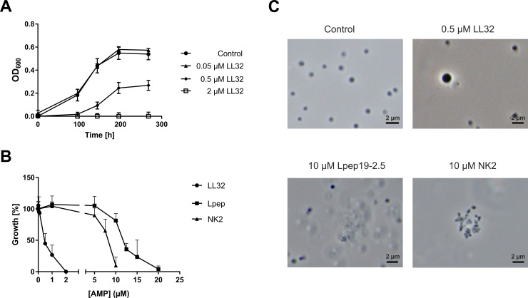
Fig 1. Growth inhibition of M. luminyensis by various AMPs.
A) 2x10⁷ cells of M. luminyensis incubated with depicted concentrations of LL32 at 37°C in 250 μl minimal medium under anaerobic conditions (see Materials and Methods). Turbidity of cultures at 600 nm (OD600) was monitored over time. Error bars represent standard deviation of three biological replicates in one experimental setup. B) 2x10⁷ cells incubated with various concentrations of the peptides LL32, Lpep 19–2.5 or NK2 at 37°C in 250 μl minimal medium. Turbidity of control cultures at 600 nm (OD600) after 200 h of growth was set to 100%. Error bars represent standard deviation of three biological replicates. C) Phase-contrast micrographs taken after 200 h of growth with the indicated concentrations of the respective added AMPs.

