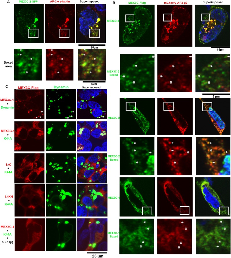
Fig 4. Subcellular localization of MEX3C in the endolysosome compartment.
A. Co-localization of MEX3C-2 (GFP-tagged) with an endogenous AP-2 α subunit. Endogenous AP-2 α was immunostained by specific antibody. The boxed area was shown at high magnification in the following row. Co-localized signals were marked by *. B. Co-localization of transiently expressed MEX3C variants (Flag-tagged, stained by anti-Flag antibody) with transiently expressed AP-2 μ2 (mCherry-tagged). The boxed area was shown at high magnification in the following row. Co-localized signals are marked by *. C. GTP binding defective dynaminK44A mutant trapped wild-type MEX3C-1, but not MEX3C-1 mutants unable to interact with AP-2, at dynaminK44A enriched foci. K44A: DynaminK44A mutant; 1ΔC: MEX3C-1ΔC, MEX3C-1 C-terminal 247AA deleted; 1ΔKH: MEX3C-1ΔKH, MEX3C-1 KH domains (AA 206–407) deleted. Shown are representative images of multiple double-positive cells. Quantitative data are presented in Fig A in S1 File.

