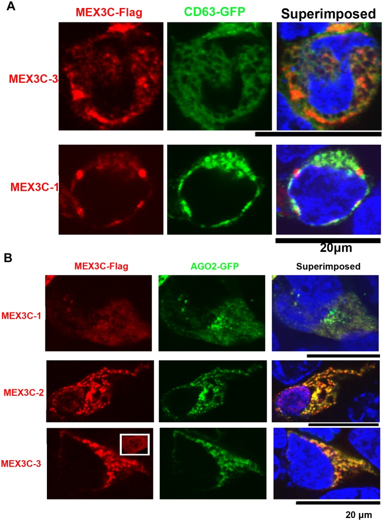
Fig 5. Co-localization of MEX3C proteins with CD63 and AGO2.
A. MEX3C proteins showed partial co-localization with CD63. B. MEX3C proteins showed a high degree of co-localization with AGO2. The boxed image shows the ubiquitous localization of singly expressed MEX3C-3. In MEX3C-3 and AGO2 double-positive cells, all MEX3C-3 protein was cytoplasmic. For A-B, MEX3C proteins were Flag-tagged; CD63 and AGO2 were GFP-tagged and their similarity to endogenous proteins was validated by respective donating investigators (see Acknowledgments for list of investigators). Shown are representative images of multiple double-positive cells. Nuclei (stained by DAPI) were pseudocolored blue.

