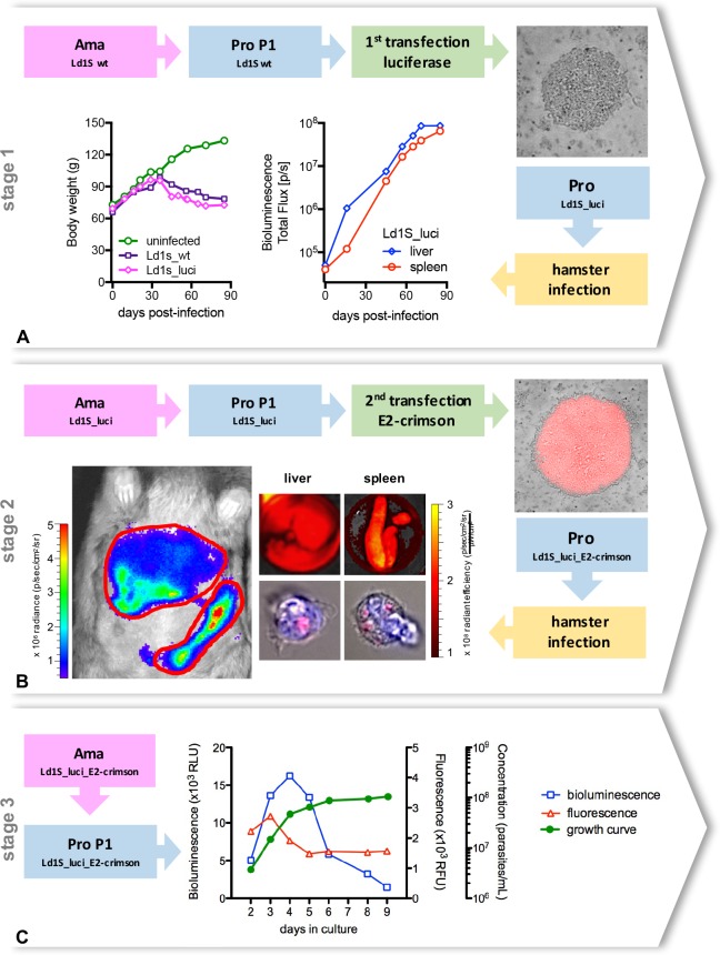
Fig 1. Generation of double transfected Leishmania donovani parasites.
(A) The first stage of the transfection aimed to generate bioluminescent parasites. Wild-type amastigotes from the Ld1S strain obtained from hamster spleen were transformed into promastigotes, then transfected with the luciferase gene and cloned in M199-agar dishes containing 150 μg/mL hygromycin. Positive colonies (top right corner) were selected, added to liquid M199 medium and promastigotes were cultivated until differentiation into metacyclic promastigotes in order to proceed with a hamster infection. Weight gain of the hamster infected with ‘Ld1S_luci’, in comparison with an uninfected hamster and with a hamster infected with wild-type amastigotes (Ld1s_wt) (bottom left). In vivo bioluminescence values in the liver and in the spleen of the hamster infected with ‘Ld1S_luci’ (bottom middle). (B) The second stage of the transfection aimed to generate fluorescent parasites. ‘Ld1S_luci’ amastigotes were obtained from the hamster spleen, transformed into promastigotes, transfected with the E2-crimson gene and cloned in M199-agar dishes containing 50 μg/mL of nourseothricin. Fluorescent colonies (top right corner) were selected, added to liquid M199 medium and promastigotes were cultivated until differentiation into metacyclic promastigotes in order to proceed with a hamster infection with ‘Ld1S_luci_E2-crimson’. Hamster in vivo bioluminescence imaging (bottom left, and S1 Fig) and simultaneous ex vivo analyses of fluorescence in the liver and in the spleen at day 90 post-infection (top middle panels). Red fluorescent parasites are also detectable in the cytoplasm of isolated hamster cells (nuclei counterstained with DAPI) (bottom middle panels). (C) ‘Ld1S_luci_E2-crimson’ amastigotes were obtained from the hamster spleen and transformed into promastigotes, considered then the first passage of promastigotes (P1). Bioluminescence (left y-axis) and fluorescence (first right y-axis) values, as well as the growth curve (second right y-axis) obtained from P1 ‘Ld1S_luci_E2-crimson’ promastigotes cultivated in vitro are shown in the graph.

