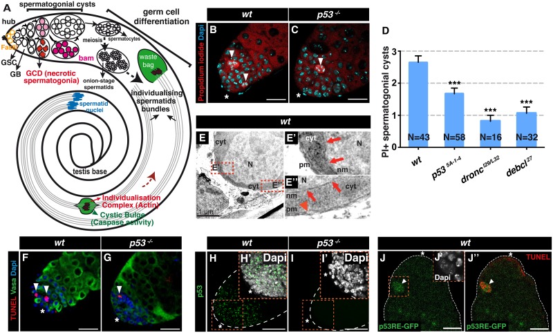
Fig 1. Drosophila p53 is required for necrotic cell death during spermatogenesis.
(A) In Drosophila testis apical tip, germ stem cells (GSCs) in contact with somatic hub cells (asterisk), which express Fasciclin III (FasIII), self-renew and generate goniablasts (GB) that produce spermatogonial cysts, some of which are eliminated by necrosis (germ cell death [GCD], in red). Increasing level of Bam (pink to magenta) induce maturation of spermatogonia into spermatocytes, which produce cysts of 64 spermatids by meiosis. Spermatids elongate with nuclei at the base of the testis (blue) and undergo individualization when F-actin investment cones form the individualization complex (IC, red). The IC moves toward the sperm tails (brown dashed arrow) within a structure known as the cystic bulge, which then forms the waste bag. Somatic cells and the seminal vesicle are omitted, and cell size is not to scale. (B, C) Propidium iodide (PI) staining of wt (B) and p53-/- (p535A-1-4, C) testes. Necrotic cells are indicated with white arrowheads. Nuclei are stained with DAPI. Scale bar, 40 μm. (D) Quantification of PI+ GCs in wt and p53-/- (p535A-1-4), dronc-/- (droncI29/L32), and debcl-/- (debcl27) mutant testes (mean ± s.e.m. of three independent experiments, N testes/genotype). ***p < 0.001 by two-tailed unpaired Student’s t-test. (E-E”) Electron micrographs of wt necrotic GC. Nucleus (N) and cytoplasm (cyt) are indicated. In the magnified views (E' and E” indicated by dashed red box in E) red arrowhead and arrows indicate plasma membrane (pm) and nuclear membrane (nm) ruptures, respectively. Scale bar in E, 1 μm. (F, G) TUNEL+ Vasa+ spermatogonial cysts (arrowheads) in wt (F) and p53-/- (p535A-1-4, G) adult Drosophila testes. TUNEL+ cells are indicated with white arrowheads. Nuclei are stained with DAPI and the hub region is indicated with a white asterisk. Scale bar, 40 μm. (H, I) p53 immunostaining of wild-type (wt) and p53-/- adult Drosophila testes. Nuclei are stained with DAPI (insets H', I'). Scale bar, 40 μm. (J-J”) GFP immunostaining (green in J and J”) of adult Drosophila testes harboring the p53RE-GFPnls reporter and co-stained for TUNEL (red in J”). A GFP+ TUNEL+ necrotic spermatogonial cyst (orange box in J) is indicated by a white arrowhead (J and J”). The hub region is indicated with a white asterisk (J and J”). Nuclei are stained with DAPI (inset J' corresponding to the orange box in J). Scale bar, 30 μm.

