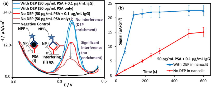
Figure 8.
(a) Anti-mouse immunoglobulin (IgG) antibodies cause false positives to the PSA immunoassay, as per the electron transfer schematics shown in the inset: (i) vs. (ii) (assay details in reference22). In absence of nDEP enrichment in the nanoslit, signals from 50 pg/mL of PSA are ~2.5-fold higher in presence of 1 µg/mL IgG, whereas with nDEP enrichment, these false positives are insignificant. (b) Signal versus time plot shows that nDEP enrichment leads to rapid enhancement in binding kinetics of PSA, with near steady-state signals after 2 minutes, whereas in absence of enrichment, the signal level does not flatten out due to false positives.

