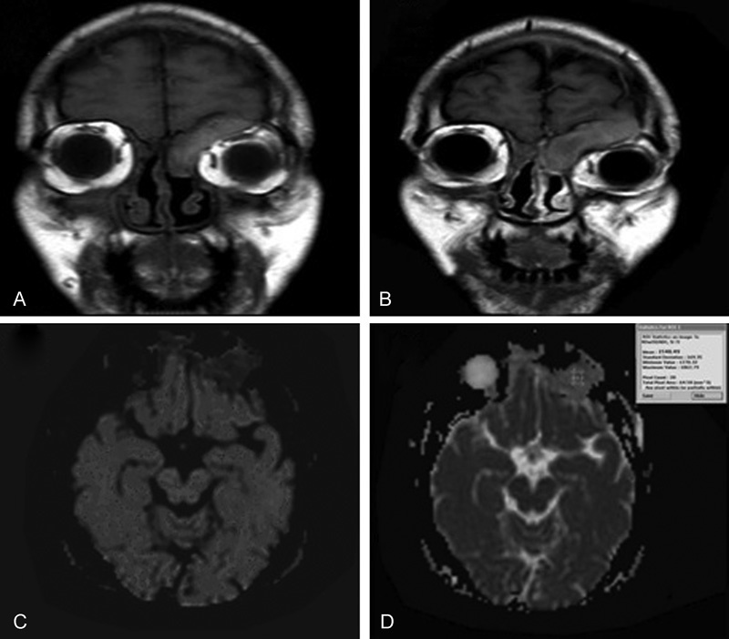
Fig. 2.

Fungal sinusitis. ( A, B ) Coronal T1WI and Coronal T1W gadolinium-enhanced MR image reveals intense enhancement of the lesion and thick uniform peripheral enhancement of left and right subperiosteal orbital abscess. ( C ) Axial DW image with b-value 1000 /mm 2 shows low signal intensity of the lesion denoting facilitated diffusion. ( D ) Axial ADC map image with b-value of 1000 second/mm 2 shows relatively high signal intensity of the lesion denoting facilitated diffusion with ADC value is 1.7 × 10⁻³ mm 2 / s. Readings suggestive of benign lesion.
