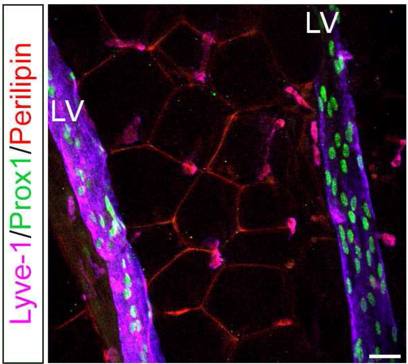
Figure 2. Presence of lymphatic vessels in fat depots of adult mice.
Visceral fat depots were dissected and subject to whole mount immunostaining with antibodies against lymphatic endothelial cells (Lyve1, Prox1) and adipocytes (Perilipin). Lymphatic vessels are seen deep inside the WAT and nearby blood vessels (not shown). LV: lymphatic vessel. Scale bar 30 uM.

