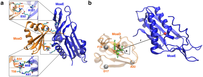
Figure 5.
Evolutionary couplings and system representation of the E. coli molybdopterin synthase subunits. (a) The crystal structure (PDB ID: 1FM061) of MoaD (orange) and MoaE (blue) with the top five scoring evolutionarily coupled pairs displayed. (b) The coordinate system used for MSM analysis of MoaD-MoaE association, where a basis set was formed using the α-carbons of three MoaD residues and the dynamics of the system were described using the coordinates of the MoaE center of mass vector projected onto the MoaD basis.

