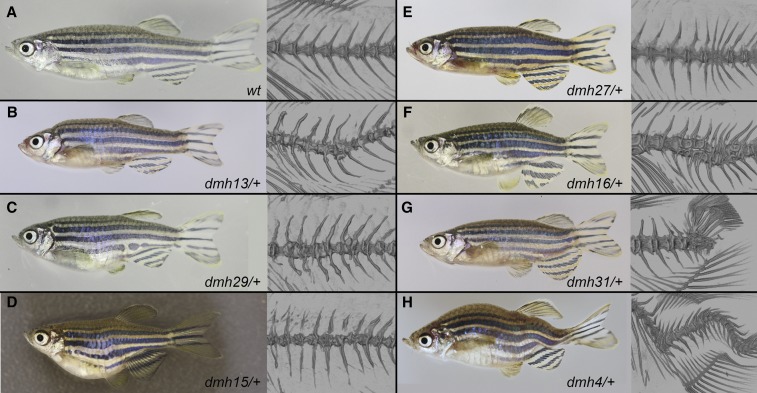
Figure 3.
Mutants with vertebral defects. Representative photographs (left) and μCT images of the vertebral column (right) of the five different subgroups of mutants with vertebral defects. (A) wild-type zebrafish, regularly patterned and shaped vertebrae, and vertebral spines. (B–D) Heterozygous dmh13, dmh29 and dmh15 mutants have a shorter trunk, and show strongly deformed vertebral bodies and spines with excessive bone formation. (E) Heterozygous dmh27 fish are also shorter than wild-type fish, but show mostly normal patterned vertebral bodies with the exception of a couple of fused vertebrae. Vertebral spines show slight changes in angle. (F) In addition to a shorter body, heterozygous dmh16 mutants have strong deformation of their vertebrae. In addition to fusion of vertebral bodies, some vertebrae are split up into multiple hemi-segments. (G) The body of dmh31 mutants seems to be proportionally normal until the region in front of the caudal peduncle. Here, vertebrae are strongly deformed, leading to a bend in the body axis. (H) Strong curvature of the spine in dorsal, ventral, and lateral direction leads to an overall shorter body length of the dmh4 mutant. The vertebral bodies seem to be mostly normally patterned.

