Figure 2.

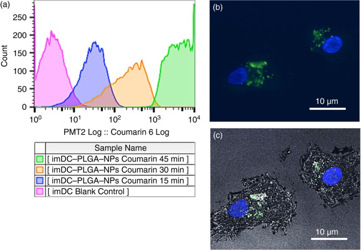
Cellular uptake and colocalization of poly(lactic‐co‐glycolic) acid nanoparticles (PLGA‐NPs) in human dendritic cells (DCs). Human immature DCs (imDCs) at 50 000/ml were cultured in 12‐well plates with 100 μg/ml of the PLGA‐NP suspension that was loaded with the fluorescent probe coumarin 6 for 15, 30 and 45 min. The cells were collected and washed three times with Hanks’ balanced salt solution to remove the uninternalized PLGA‐NPs. (a) Flow cytometry was used to analyse the kinetics of the internalization of the PLGA‐NPs by the imDCs. (b) Human imDCs incubated with PLGA‐NPs for 45 min were analysed using a confocal microscope (Leica TCS SP2). Confocal images of imDCs loaded with PLGA‐NPs; the DAPI and FITC channels were overlaid. (c) Confocal images of human imDCs loaded with PLGA‐NPs; the DAPI, FITC and reflection channels were overlaid. Scale bar = 10 μm. The flow cytometry analysis and confocal image stack scanning were independently repeated three times, respectively; each condition was performed in triplicate. [Colour figure can be viewed at wileyonlinelibrary.com]
