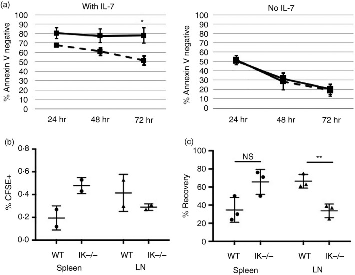
Figure 1.

Decreased survival and altered homing of Ikaros null T cells. (a) Cell viability assay for purified wild‐type (solid) and Ikaros null (IK −/−) (dashed) CD4 T cells in culture with or without 10 ng/ml of exogenous interleukin‐7 (IL‐7) measured as per cent of cells that stained negative for Annexin V. Data shown are a compilation from four independent experiments (*P = 0·00684). (b) Purified CD4 T cells from wild‐type and Ikaros null spleens were loaded with CFSE and adoptively transferred into two separate wild‐type mice. Percent (%)CFSE + cells in the spleen and lymph nodes are shown. (c) Differentially labelled wild‐type (Efluor 450) and Ikaros null (CFSE) CD4 T cells were adoptively transferred in a 1 : 1 ratio into the same wild‐type mouse. (NS = Not significant, P = 0·0501; **P = ·0062). % Recovery = Individual percentage of wild‐type (Efluor 450) or Ikaros null (CFSE) cells in the spleen or lymph node/ total percentage of labelled (Efluor 450 + CFSE) cells in the spleen or lymph node. Error bars are representative of ± SEM.
