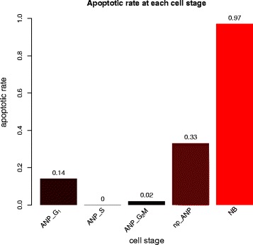
Fig. 8.

Estimated apoptosis rate at each cell stage that yields best fit to data. The bar graphs show estimated apoptotic rates at each cell stage through the early stages of the hippocampal neurogenic cascade in a 1-month old mouse. The apoptosis is highest among neuroblasts (NBs), followed by non-proliferating ANPs (np-ANP). This is in agreement with experimental data, indicating that model fit is good
