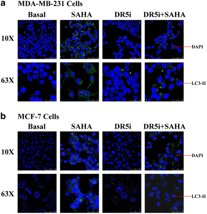
Figure 4.

LC3-II immuno-fluorescence assay induced by SAHA and TRAIL DR5. TRAIL DR5 transfected cells of MDA-MB-231 (a) or MCF-7 (b) were incubated with SAHA in 6-well plate. The cells were fixed with 4% formaldehyde at room temperature and blocked with buffer solution for 2 h at room temperature. LC3-II antibody solution was added to incubate with cells overnight at 4 °C. A DyLight 488 fluoresence antibody (1 : 200 dilution) was used for 1 h incubation and nuclei were counterstained with DAPI dye for another 10 min. The cells were imaged and autophagy signals were visualized by Leica DMI6000 B microscope with ×10 (scale bars indicate 50 μm) and ×63 (scale bars indicate 25 μm) magnification.
