
Fig. 5.
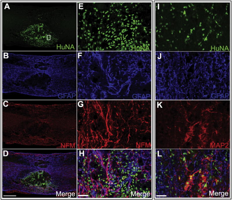
A2B5+ sorted cells derived from patient U-iPSCs give rise to neurons and astrocytes after being grafted to SCI mouse model. FACS purified A2B5+ cells are grafted into the contused thoracic spinal cord of adult mice. Eight weeks after transplantation, grafted human cells survive and integrate into the injured spinal cord as shown by the human nuclei staining (HuNA) (A, E, I). Some have matured to neurons and start to express neuronal marker NFM and MAP2 (C, H, K) some differentiate into GFAP+ astrocytes (B, F, J). Bar, A–D, 500 μm; E–H and I–L, 20 μm. E–H represent higher magnification images of the boxed areas in A–D.