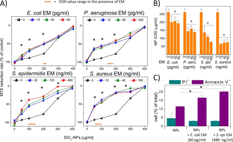
FIG 1.
DC cytotoxicity induced by bacterial EMs and silica NPs. (A) DCs were incubated for 24 h with different NP concentrations in the presence or absence of the indicated EMs. The supernatants were aspirated, and cells were incubated with MTS solution until color development. The percent MTS reduction was calculated with respect to the amount for nontreated cells (control). Data are the means ± SEs (n = 3, run in triplicate). *, P < 0.05 with respect to cells not treated with EMs. (B) IC50 (SiO2 NP doses resulting in 50% of the maximal cytotoxic effect, as measured by the MTS assay) values were plotted against EM concentrations. Data are means ± SEs (n = 3, run in triplicate). *, P < 0.05 with respect to cells treated with NPs alone. (C) DCs, treated as described above, were stained with annexin V-FITC and propidium iodide (PI) and subsequently subjected to FACS analysis. Bars represent the mean percentage ± SE (n = 3) for assays run in triplicate of PI-positive or annexin V-positive cells. *, P < 0.05 with respect to cells treated with NPs alone.

