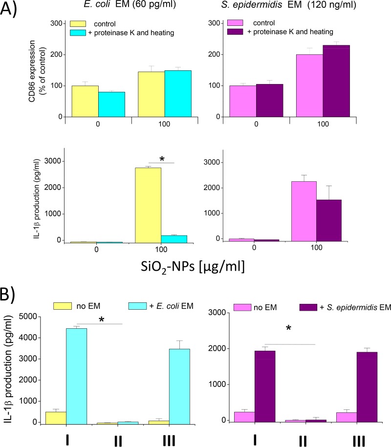
FIG 9.
Dependence of NP and EM synergy on proteinaceous PAMPs and on an association with bacterial factors on the surface of NPs. (A) DCs were treated (for 24 h at 37°C) with EMs or with EMs that had been incubated for 1 h at 50°C with 100 μg/ml proteinase K and then for 5 min at 95°C in order to degrade proteins; the cells were then analyzed for CD86 expression (top), and the amount of IL-1β in the culture supernatant was measured (bottom); data are from one representative experiment of three experiments conducted and represent means ± SEs. *, P < 0.05 with respect to cells not treated with proteinase K. (B) NPs (100 μg/ml) were incubated with the indicated EM concentrations for 20 min at 37°C in RPMI 1640 containing 10% FBS. The NPs were then centrifuged for 30 min at 13,000 rpm, and the supernatant was recovered. The NPs were washed three times with PBS, and finally, some NPs were resuspended in fresh medium and other NPs were resuspended with the recovered supernatant. Then, the NPs were used to treat DCs for 24 h at 37°C and IL-1β production was measured by ELISA. Data are from one representative experiment of three experiments conducted and represent means ± SEs. *, P < 0.05. I, NPs coincubated with the indicated medium and DCs; II, NPs preincubated with the indicated medium, pelleted, and incubated with DCs after resuspension in new medium without EM; III, NPs preincubated with the indicated medium, pelleted, and incubated with DCs after resuspension in their corresponding supernatants.

