
Fig. 5.
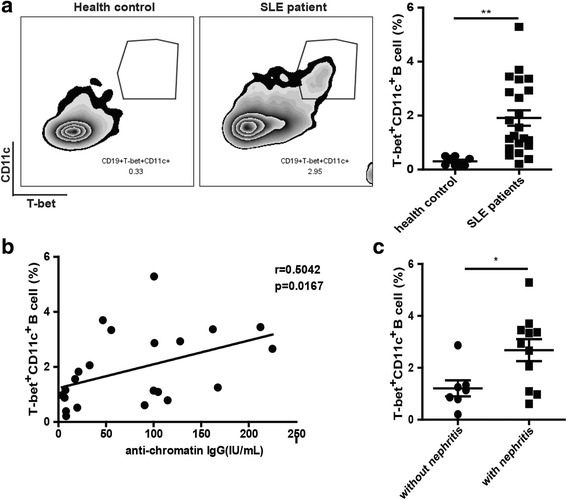
The percentage of T-bet+CD11c+CD19+ B cells was elevated and associated with antichromatin autoantibody in SLE patients. a Flow cytometric analysis of T-bet+CD11c+CD19+ B cells in peripheral blood from systemic lupus erythematous (SLE) patients (n = 22) and healthy donors (n = 10). b Correlation between the percentage of T-bet+CD11c+CD19+ B cells and the level of antichromatin autoantibody. c The percentage of T-bet+CD11c+CD19+ B cells exhibited an increasing trend in patients with lupus nephritis (n = 14) relative to patients with no history of lupus nephritis (n = 8). In (a) and (c) values are shown as the mean ± SD. *P < 0.05; **P < 0.01. Symbols (filled circles and squares) represent individual subjects. IgG immunoglobulin G