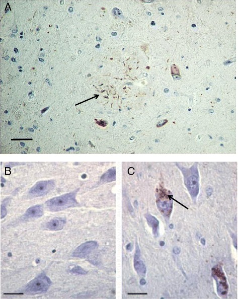
Fig. 2.

Phosphorylated PKR immunostaining in AD and control brains. a AD brain PKR staining is localized to neuritic dendrites surrounding an amyloid plaque, as well as in the cytosol of neighboring neurons (bar = 60 μm). The arrow indicates dystrophic neurites positive for pPKR staining. b No neuronal staining is depicted in a control brain (bar = 20 μm). c AD brain PKR immunostaining is seen in neuronal cytosolic vacuoles (bar = 20 μm). The arrow indicates pPKR accumulation in neurons from an AD brain. Original figure from authors
