
Figure 1.
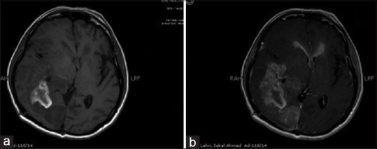
MRI Brain T1W axial non-contrast enhanced (a) and contrast-enhanced image (b) showing heterogeneously enhancing space occupying lesion with distant enhancement of walls of the bilateral frontal horns
Official websites use .gov
A
.gov website belongs to an official
government organization in the United States.
Secure .gov websites use HTTPS
A lock (
) or https:// means you've safely
connected to the .gov website. Share sensitive
information only on official, secure websites.
MRI Brain T1W axial non-contrast enhanced (a) and contrast-enhanced image (b) showing heterogeneously enhancing space occupying lesion with distant enhancement of walls of the bilateral frontal horns