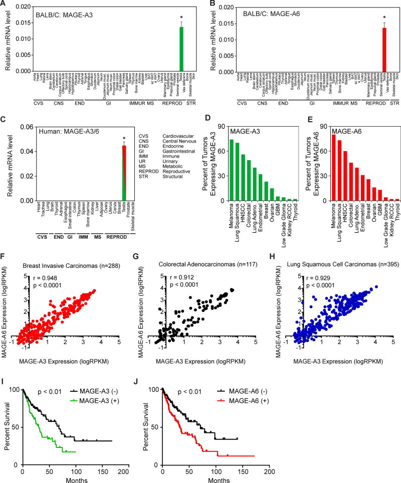
Figure 1. MAGE-A3 and MAGE-A6 are normally restricted to expression in the testis, but are aberrantly expressed in cancer and predict poor patient prognosis.
(A–B) RT-QPCR analysis (n=3) of the normalized expression of mouse MAGE-A3 (A) and MAGE-A6 (B) in the indicated tissues from BALB/C mice.
(C) RT-QPCR analysis (n=3) of the normalized expression of human MAGE-A3/6 (one primer set detects both) in the indicated human tissues.
(D–E) Percent of patient tumors expressing MAGE-A3 (D) and MAGE-A6 (E) is shown.
(F–H) MAGE-A3 and MAGE-A6 are co-expressed in breast invasive carcinomas (F), colorectal adenocarcinomas (G), and lung squamous cell carcinomas (H).
(I–J) Expression of MAGE-A3 (C) or MAGE-A6 (D) in patients with lung squamous carcinomas correlates with poor overall survival.
Data are represented as the mean +SD. Asterisks indicates p<0.05. See also Figure S1 and Table S1.

