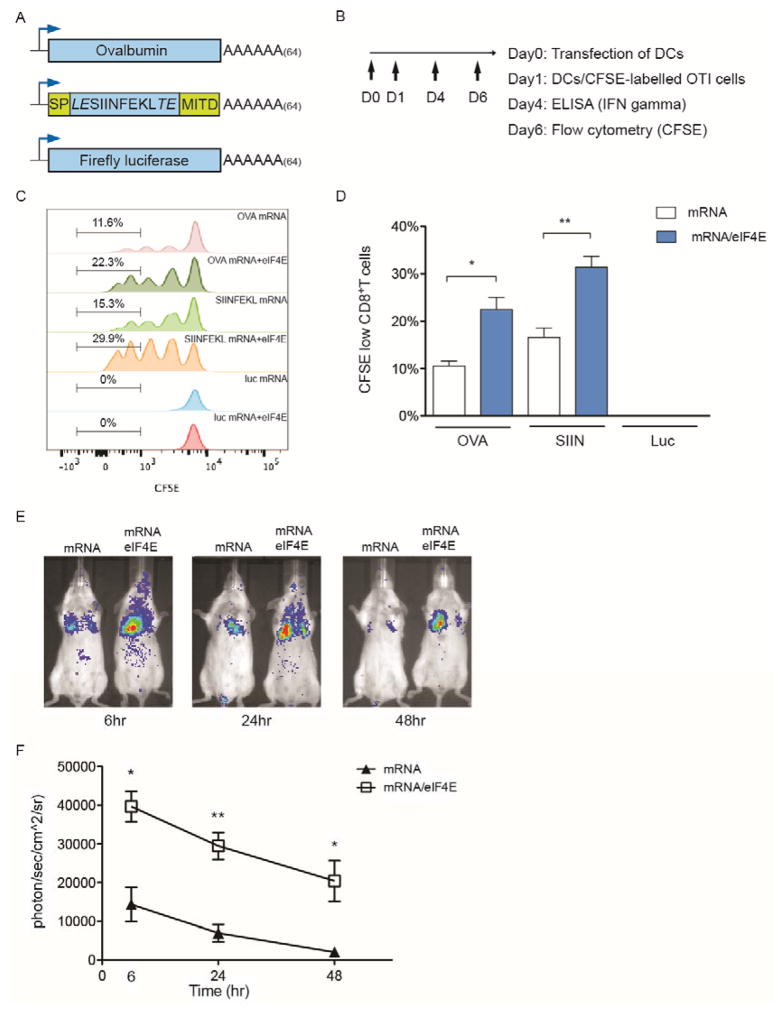
Figure 6. Delivery of mRNA/eIF4E nanoplexes enhances mRNA expression ex vivo and in vivo.
(A) Schematics of different mRNA templates used in the study: Full length OVA; SIINFEKL peptide flanked by a MHC class I signal peptide fragment (78 bp) and a MHC class I trafficking signal (MITD, 168 bp); Luciferase mRNA. (B) Procedures for ex vivo antigen presentation. (C) Representative histograms of proliferation of OTI CD8 T cells on day 6 in response to antigen presentation by DCs. DCs were transfected with antigen mRNA alone or mRNA/eIF4E nanoplexes on day 0 through N5 (PEH). (D) Percentage of activated CD8 T cells through CFSE labeling based on the same gating as in (C). The activation of CD8 T cells was identified by the decrease of CFSE fluorescence intensity via flow cytometer. (E) Representative bioluminescence imaging (BLI) at 6, 24 and 48 hr after the tail vein injection of luciferase mRNA or mRNA/eIF4E packaged with the polyamine. (F) Quantification of luciferase expression in lungs of Balb/c mice via BLI. Data represent the mean ± SEM (n=3). *, P<0.05. **, P<0.01.

