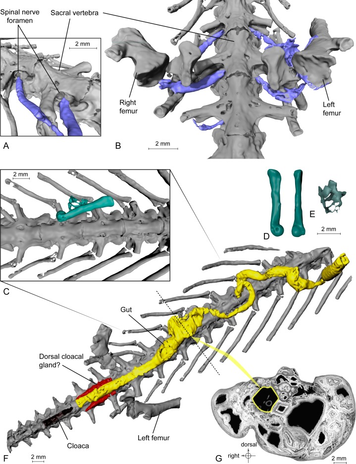
Figure 3. Exceptional preservation of nerves, digestive tract and stomachal content.
(A and B) 3D reconstructions of the pelvic section of MNHN.F.QU17755, in laterodorsal (A) and ventral (B) views. The lumbosacral plexus (in blue) is partly preserved. Nerves exit the last trunk, the sacral and the first caudosacral vertebrae through the spinal nerve foramina. (C) Preserved bones of an anuran frog (ranoid?), in green, inside the digestive tract (not shown, to better reveal its content; see Fig. 3F) of MNHN.F.QU17755. (D) Anuran humerus found inside digestive tract of MNHN.F.QU17755, in lateral and ventral views. (E) Anuran vertebrae found inside digestive tract of MNHN.F.QU17755. The centrum is very thin; the holes may represent segmentation artifacts. (F) 3D reconstruction of MNHN.F.QU17755 in ventral view, showing the nearly complete digestive tract. The caudal end is very close to the cloaca, and is bordered near the pelvic girdle by presumed dorsal cloacal glands (see Fig. 4A). The dotted line represents the position of the virtual section illustrated in Fig. 3G. (G) Virtual section of the trunk, showing the digestive tract (in yellow) and its content (frog bones).

