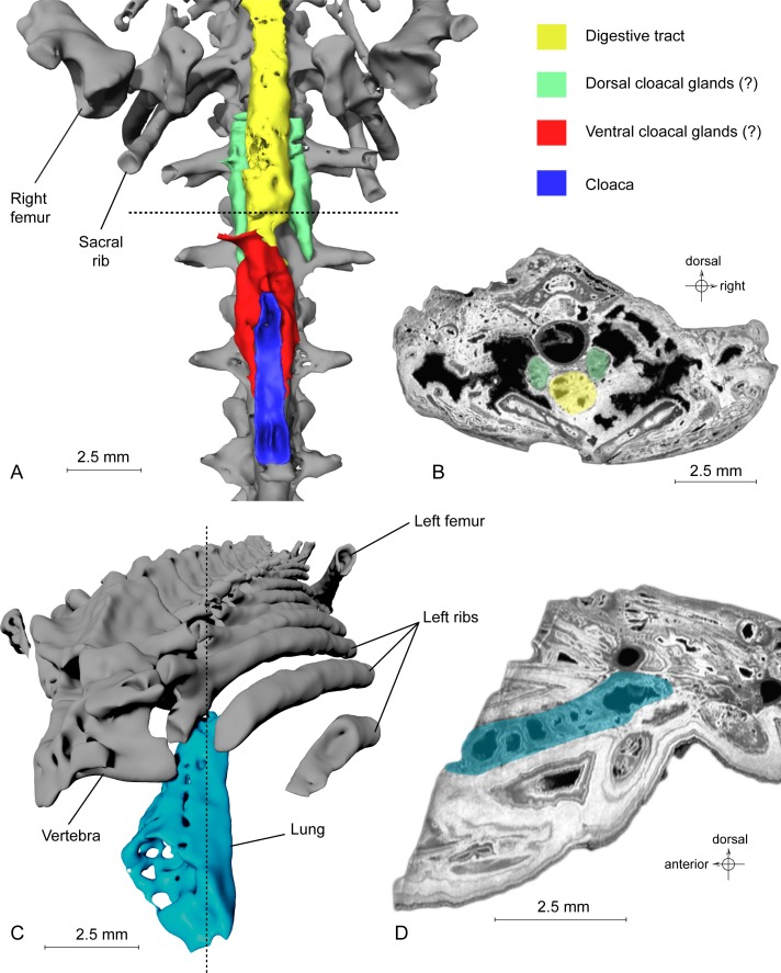
Figure 4. Exceptional preservation of cloacal glands (?) and lung.
(A) 3D reconstruction of supposed dorsal and ventral cloacal glands, in ventral view, under the two ischia (not shown). The dorsal cloacal glands are located between the first and second caudosacral vertebrae and the digestive tract (see Fig. 4B). The ventral cloacal glands are located under the digestive tract and anterodorsal to the cloaca. The dotted line represents the position of the virtual section illustrated in Fig. 4B. (B) Virtual section of the pelvic girdle, illustrating the digestive tract and the dorsal cloacal glands, between a caudal vertebra and the two ischia. (C) 3D reconstruction of the incomplete lung (in blue), inside the specimen MNHN.F.QU17755, in oblique anterior view. It is located lateroventrally to the trunk vertebrae, in the anteriormost preserved part of the fossil. The dotted line represents the position of the tomogram illustrated in Fig. 4D. (D) Virtual section of the anteriormost preserved part of MNHN.F.QU17755, showing the inside of the lung in lateral view.

