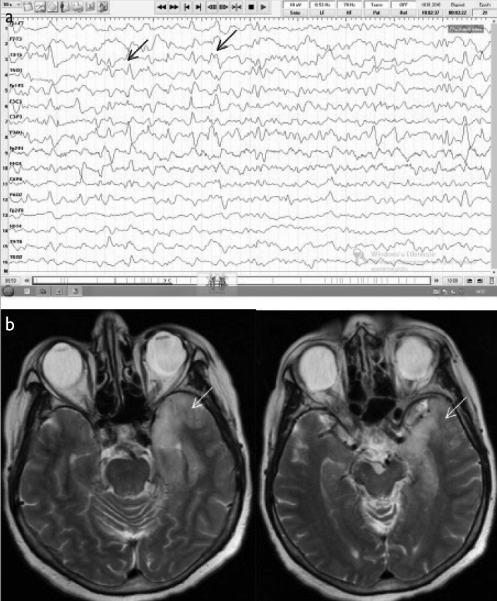
Figure 1. a, b.

A 67-year-old female patient presenting with altered behavior was eventually diagnosed with PCR-confirmed herpes encephalitis. (a) Initial EEG (double banana montage) showed diffuse bioelectrical disorganization of 6–7 Hz frequency prominent on the left as well as repetition of sharp wave activity every 0.5 s (black arrows). (b) The cranial MRI (T2) revealed left temporal hyperintensity (white arrows).
