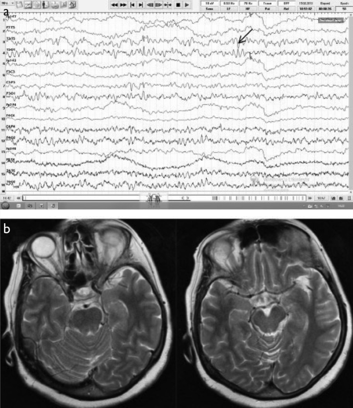
Figure 2. a, b.

A 63-year-old female patient presenting with nonsensible speech was eventually diagnosed with PCR-negative possible viral encephalitis. (a) Initial EEG (double banana montage) showed diffuse bioelectrical disorganization on both temporal hemispheres that was more prominent on the left and temporoparietal neuronal hyperexcitability (arrow). (b) The cranial MRI (T2) was normal.
