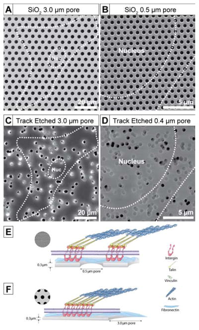
Figure 1.
Scanning electron microscopy (SEM) images of 300 nm thick SiO2 membranes with (A) 3.0 μm and (B) 0.5 μm diameter pores; Greiner Bio-One Thincert® track etched high-porosity membranes with (C) 3.0 μm and d) 0.4 μm diameter pores. Dotted white outline shows the size of a typical cell and its nucleus. Illustrations (E) and (F) show potential focal adhesion formations on 0.5 μm and 3.0 μm diameter pore SiO2 membranes.

