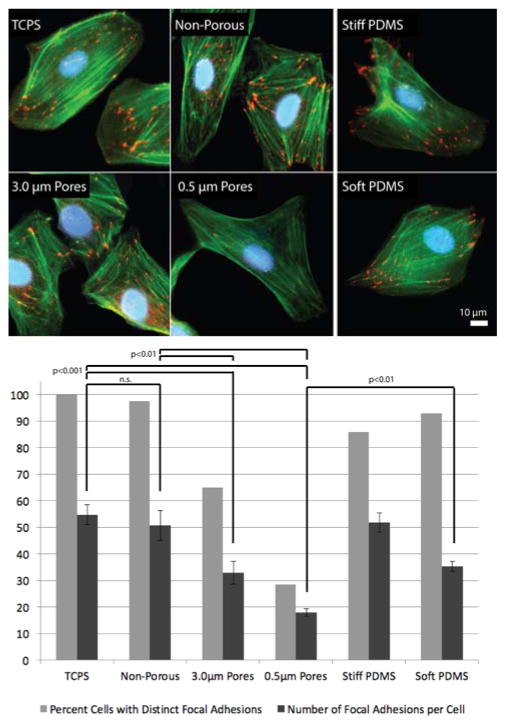
Figure 2.
Representative images of endothelial focal adhesions after 24 hours on TCPS, SiO2 membranes, Stiff (2 MPa) PDMS and Soft (5 kPA) PDMS substrates. Cells were stained for nuclei (DAPI, blue), F-actin (phalloidin, green), and focal adhesions (anti-vinculin, red). Bottom: Quantification of focal adhesion formation after 24 hours of culture. Formation of distinct focal adhesions was quantified for all substrates - percent of cells with distinct focal adhesions and number of focal adhesions per cell (n > 20 for each substrate; mean ± standard deviation; one-way ANOVA with a Tukey post hoc analysis).

