Table 1.
Recent advances of nanoscaffold strategies to induce blood vessel regeneration.
| Materials | Nanostructures/cellular morphology | Study model | Outcome | References |
|---|---|---|---|---|
| Nanoscale-structured vascular scaffolds | ||||
| PGA/gelatin nanofiber (87.72 ± 23.34 nm) |

|
In vitro | Significantly higher mechanical properties on PGA/gelatin; enhanced EC and SMC growth on PGA with 10% and 30% gelatin, respectively | Hajiali et al.[18] |
| Self-assembled peptide gel (ligated CQ11*G-thioester, 11–13nm fibrils) |

|
In vitro | Greater EC proliferation and CD31expression by the ligated peptide | Jung et al. [24] |
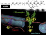
| Heparin-binding peptide amphiphile (HBPA) nanofibers |
|
In vitro | HBPA nanofibers induced superior tubule-like interconnected networks by EC than scrambled PA | Rajangam et al. [23] |
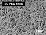
| PEGylated fibrin (200–400 nm) |
|
In vitro/in vivo | Tunable nanofiber diameter and storage modulus based on chemical modification | Zhang et al. [15] |
| Nanoscale surface-modified vascular scaffold | ||||
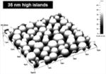
| Polymer demixed nano-islands (13–95 nm) |
|
In vitro | More spread EC morphology depending on altered nanotopography | Dalby et al. [32] |
| PEG-polyurethane substrates (1.5–40nm) |

|
In vitro | Enhanced HUVEC proliferation on higher levels of nanorough surfaces modified with mixed different PEGs | Chung et al. [36] |
| Nanoscale-patterned titanium surfaces (750 nm-100μm space between grooves) |

|
In vitro | Significantly enhanced RAEC density at 4 h/1 day on the nanopatterned surface | Lu et al. [30] |
| Nanopatterned PMMA and PDMS (350 nm linewidth, 700 nm pitch, 350 nm depth) |

|
In vitro | Decreased SMC growth on nano-patterned PMMA/PDMS compared to flat surfaces; enhanced MTOC polarization on nano-patterned surfaces | Yim et al. [34] |
| Nanostructured PLGA |

|
In vitro | Significantly increased surface roughness in NaOH-treated and cast PLGA; increased SMC growth but decreased EC growth on treated PLGA | Miller et al. [35] |
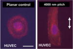
| Nanogrooves in polyurethane (200–2000 nm) |
|
In vitro | Organization and alignment of ECs in nanogrooves; differential response based on anatomic origin of ECs | Liliensiek et al. [39] |
| Titanium stent coated with Rosette nanotube-lysine side chain (3.5 nm diameter) |
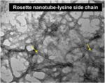
|
In vitro | Increased EC adherence and proliferation | Fine et al. [31] |
| Nano-patterned PLGA microvascular scaffolds |
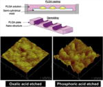
|
In vitro | More controlled pattern of EC residence and growth on the 20 nm nanosurface than the 80 nm | Wang et al. [33] |
All figures reprinted or adapted with permission.
