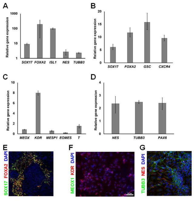
Figure 5.
Embryoid body (EB) formation and multilineage differentiation for hPSCs expanded in VN+HSA+UV microcarrier cultures for 5 continuous passages. (A) Cells within EBs displayed markers of DE, MS and NE analyzed by qPCR. Expression was normalized to that of undifferentiated cells with ACTB as the housekeeping gene. Quantitative PCR analysis of hPSCs expanded for 5 passages in microcarrier cultures and subjected to directed differentiation toward (B) DE, (C) MS and (D) NE and the corresponding immunocytochemistry results (E–G). The expression results in (B–D) was normalized to that of hPSCs cultured in basal media without the differentiation factors.

