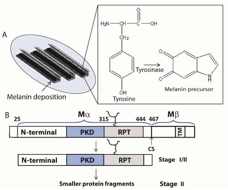
Figure 2.
(A) Schematic representation showing stage III/IV melanosomes. Melanin is depicted as black polymer deposits on preformed fibrils (left). Chemical transformation of L-tyrosine by tyrosinase to the melanin precursor, indole-5,6-quinone (right). (B) Stage I contains full-length Pmel17, which undergoes membrane cleavage, liberating the Mα from the Mβ transmembrane fragment. Further proteolytic processing of Mα (Stage II) generates smaller fragments containing at least two domains, including RPT, which contributes to the fibrillar matrix.

