Figure 2.
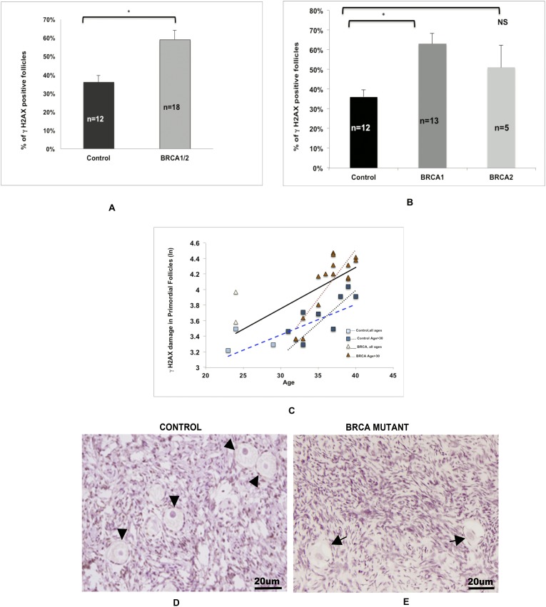
Effect of BRCA mutations on DNA damage in primordial follicle oocytes. (A) The proportion of γH2AX-positive primordial follicle oocytes was higher in the ovaries of BMCs than in the ovaries of controls (59% ± 5% vs 36% ± 3%; P = 0.0005). (B) Compared with controls (36% ± 3%), a higher likelihood of DNA DSBs was observed in the primordial follicle oocytes of the BRCA1 subgroup (63% ± 5%; P = 0.0003) but not the BRCA2 subgroup (51% ± 11%; P = 0.24). The error bars represent standard error of the mean. Statistical significance is denoted by asterisks (*). (C) Comparison of the linear regression curves by multivariate analysis indicates that there was a faster accumulation of DNA breaks (R2 = 0.57; P = 0.002) in primordial follicle oocytes of BMC ovaries (slope = 0.0530) than in control ovaries (slope = 0.0390). This difference between the BMCs (slope = 0.1263) and controls (slope = 0.081) became more distinct when the analysis was limited to those aged >30 years. The squares represent the control group, and the triangles represent the BMCs. Dotted lines represent the analyses that were limited to age >30 years in each group. Refer to the text for linear regression results. Photomicrographic representation of γH2AX expression in (D) controls and (E) BMCs. (D) Arrowheads show the γH2AX− follicles in controls. (E) Arrows mark the γH2AX+ follicles in BMCs.

