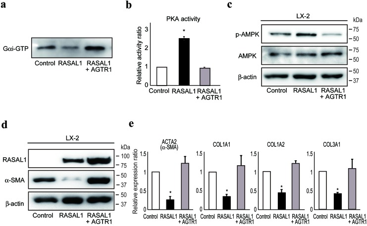
Figure 5. RASAL1 interacts with AGTR1 and regulates Gαi function.
a., Gαi activity in control (Control), RASAL1-expressing (RASAL1), and RASAL1- and AGTR1-expressing (RASAL1 + AGTR1) LX2 cells subjected to GTP-bound active Gαi pull-down assay. A representative result from five independent experiments is shown. b., PKA activity in control (Control), RASAL1-expressing (RASAL1), and RASAL1- and AGTR1-expressing (RASAL1 + AGTR1) LX2 cells measured by ELISA using cell lysates and phosphorylation-specific antibodies against PKA substrate peptide. Data are means ± SD of three independent experiments. *, p < 0.05 (t-test). c., d., The levels of phosphorylated AMPK (c) and α-SMA protein (d) in control, RASAL1-expressing, and RASAL1- and AGTR1-expressing LX2 cells were determined by Western blotting. e., Expression levels of fibrosis-related gene transcripts were assessed by quantitative RT-PCR. Values are the mRNA expression levels in RASAL1-expressing cells and RASAL1- and AGTR1-expressing cells relative to those in control LX2 cells. Data are means ± SD of three independent experiments. *, p < 0.05 (t-test).

