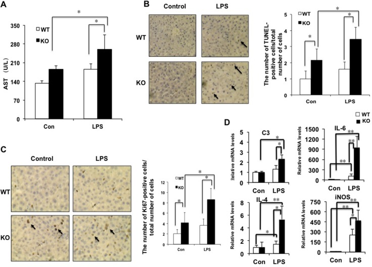
Figure 3. The deficiency of miR-149* in mouse liver is more sensitive to LPS-induced liver injury.
8-week-old WT and miR-149*−/− (KO) mice were treated with LPS (10mg/Kg body weight) for 16 hours. Then the mouse livers and blood were collected for further analysis. (A) AST levels of wild-type (WT) and miR-149*−/− (KO) mice after LPS treatment (n = 5–6). Con, control; LPS, LPS-treated groups. P < 0.05. (B) Representative TUNEL staining of sections from WT and miR-149*−/− livers (magnification X200) and statistical analysis of the number of TUNEL-positive cells per total number of cells. The number of cells in at least 20 microscopic fields was counted. P < 0.05 (n = 5–6). Black arrows, TUNEL-positive cells. (C) Representative Ki67 staining of sections from WT and miR-149*−/− livers (KO) (magnification ×200) and statistical analysis of the number of Ki67-positive cells per total number of cells. The number of cells in at least 20 microscopic fields was counted. *P < 0.05. Black arrows, Ki67-positive cells. (D) Quantitative real-time PCR (qRT-PCR) analysis of the expression of proinflammatory genes in livers from WT or miR-149*−/− (KO) mice (n = 5–6). *P < 0.05 and **P < 0.05.

