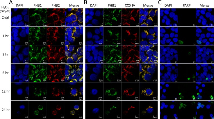
Figure 4. PHB1 and PHB2 co-localize to mitochondria of Kit225 during oxidative stress.
Kit225 cells were treated with 500 µM H2O2 for 0, 1, 3, 6, 12 or 24 hr. Cells were cytocentrifuged onto glass slides and subjected to analysis by immunofluorescent confocal microscopy. (A) PHB subcellular localization was determined by staining the nucleus with DAPI; PHB1-Alexa488 and PHB2-Cy3 and overlay (B) Nuclear staining with DAPI, PHB1-Alexa488, COX IV-Cy3 and overlay. (C) Nuclear staining with DAPI, cleaved PARP-Alexa488 and overlay were detected in Kit225 cells using immunofluorescent confocal microscopy.

