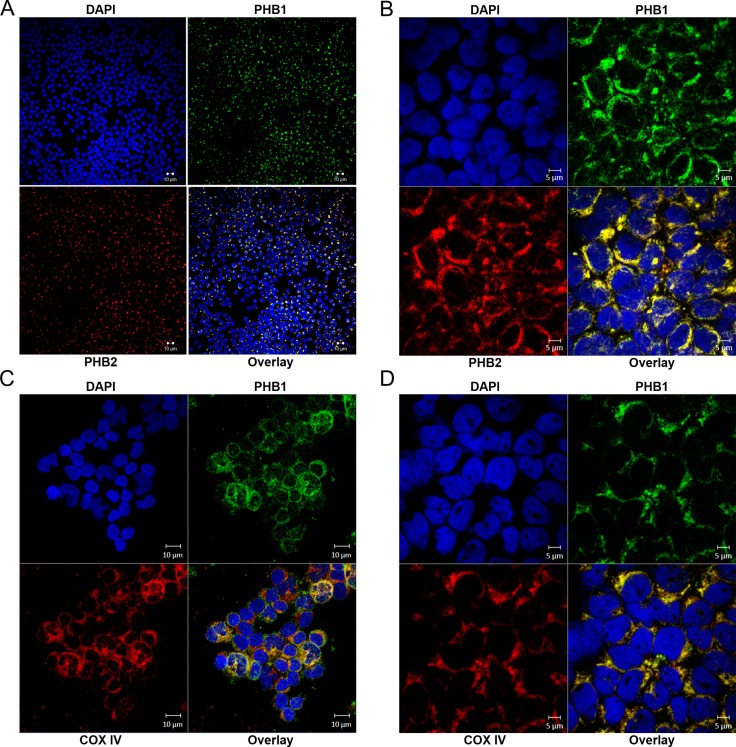
Figure 6. PHB1/PHB2 co-localize to peri-nuclear regions and mitochondria in tumor cells obtained from patients diagnosed with hematologic malignancies.
Samples were cytocentrifuged onto glass slides and subjected to analysis by immunofluorescent confocal microscopy. Immunofluorescent confocal microscopy displaying DAPI (upper left panels); PHB1-Cy2 (upper right panels), PHB2-Cy3 or COX IV-Cy3 (lower left panels) and overlay (lower right panels) in (A and C) T-ALL and (B and D) CML patient samples.

