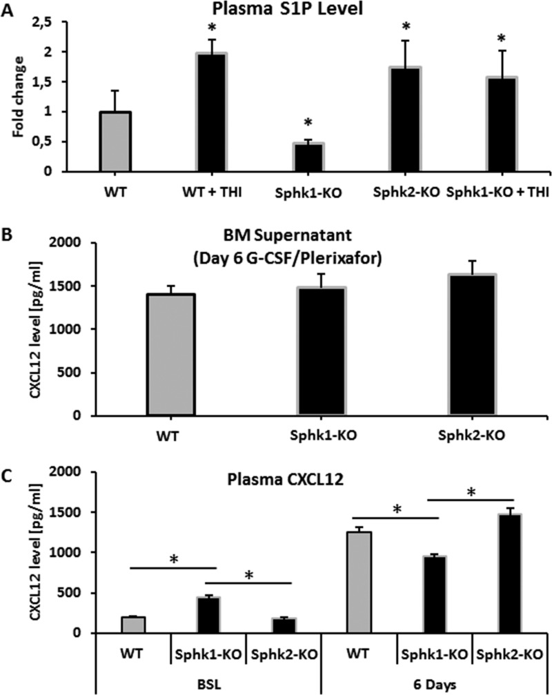
Figure 2. Measurements of S1P and CXCL12 levels.
Panel (A) Steady-state S1P plasma level in WT, Sphk1-KO, Sphk2-KO, and Sphk1-KO mice exposed to THI. Panel (B) CXCL12 concentration in conditioned media from BM after 6 days of G-CSF mobilization in Sphk1-KO and Sphk2-KO animals. Panel (C) Left: the plasma level of CXCL12 in Sphk1-KO and Sphk2-KO animals under steady-state conditions. Right: the plasma level of CXCL12 in Sphk1-KO and Sphk2-KO animals mobilized for 6 days with G-CSF. Results from two separate experiments are pooled together.

