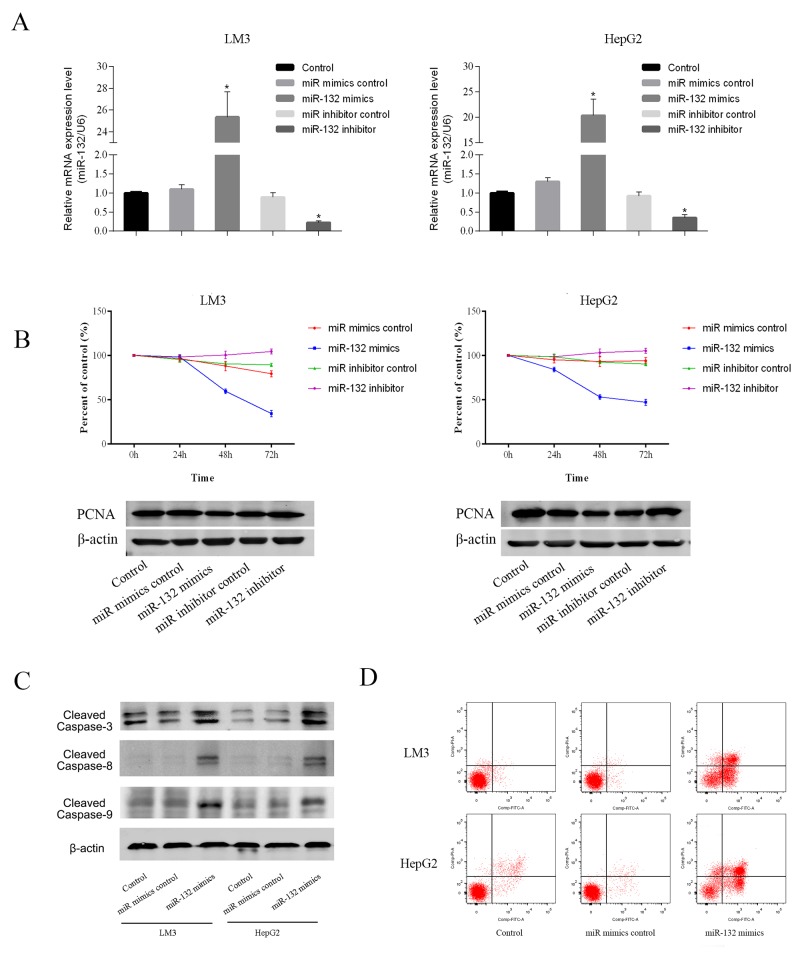
Figure 2. Effect of miR-132 on cell proliferation and apoptosis.
(A) The relative mRNA levels of miR-132 were determined by RT-PCR (n = 3, **p < 0.05 for miR-132 mimics or inhibitor vs. miR control). (B) LM3 and HepG2 cells were transfected with miR-132 mimics (50 nm) or inhibitor (100 nm). The CCK8 kit was used to monitor cell proliferation (n = 3). The protein expression of PCNA was measured by western blotting. (C) Apoptosis of LM3 and HepG2 cells was determined by flow cytometry and the protein expression of cleaved caspases was measured by western blotting.

