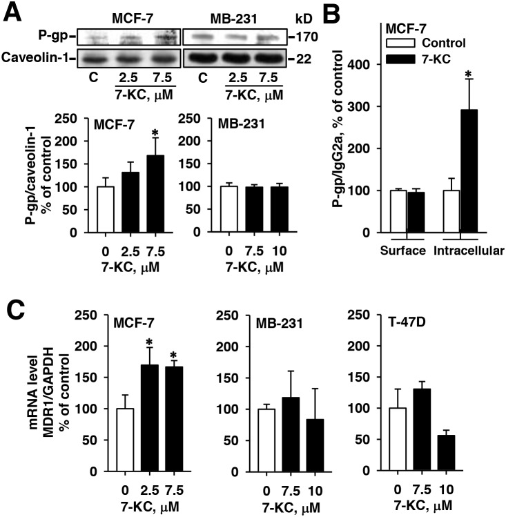
Figure 4. Alterations of protein and mRNA levels of P-glycoprotein (P-gp) in breast cancer cells after 48-h exposure to 7-ketocholesterol (7-KC).
The levels of MDR1 gene-encoded P-gp protein was analyzed by immunoblotting analysis of crude membrane proteins (50 μg protein/well) (A) and by flow cytometry using PE-UIC2 antibody (B) as described in the Methods. The protein level of caveolin-1 was determined as the internal control. The band and fluorescence intensities of immune-reacted P-gp were normalized with the respective intensities of the internal controls, caveolin-1 and IgG2a. The results are presented as means ± SD of three and four independent experiments in the analyses of immunoblotting and flow cytometry, respectively. *p < 0.05. Panel (C) shows the changes of MDR1 mRNA analyzed by a real-time polymerase chain reaction. mRNA levels of MDR1 were normalized to the level of glyceraldehyde 3-phosphate dehydrogenase (GAPDH). The results are presented as means ± SD of three independent experiments. *p < 0.05, compared to the vehicle control.

