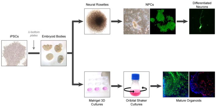
Fig. 4.
Schematic of systems for culturing patient-derived cerebral tissues. iPSCs are aggregated into embryoid bodies to allow for deposition of matrix and the formation of dense tissues. The generation of differentiated neurons involves the transfer of embryoid bodies to adhesive plates to allow for neural rosette formation. Neural rosettes are dissociated and differentiated into neural progenitor cells (NPCs), which can be further differentiated into various types of post-mitotic neurons. Representative cultured NPCs and post-mitotic neuron express TUJ1 (green). The generation of cerebral organoids involves the transfer of embryoid bodies to Matrigel, and then neural induction in stationary culture. After sufficient expansion of the resulting neuroepithelium, samples are transferred to a rotating suspension culture to promote nutrient diffusion and further organoid maturation. Mature organoids are subsequently collected for molecular and cellular analyses. Representative organoids express VIMENTIN (green) and FOXG1 (red). Nuclei are counterstained with DAPI (blue).

