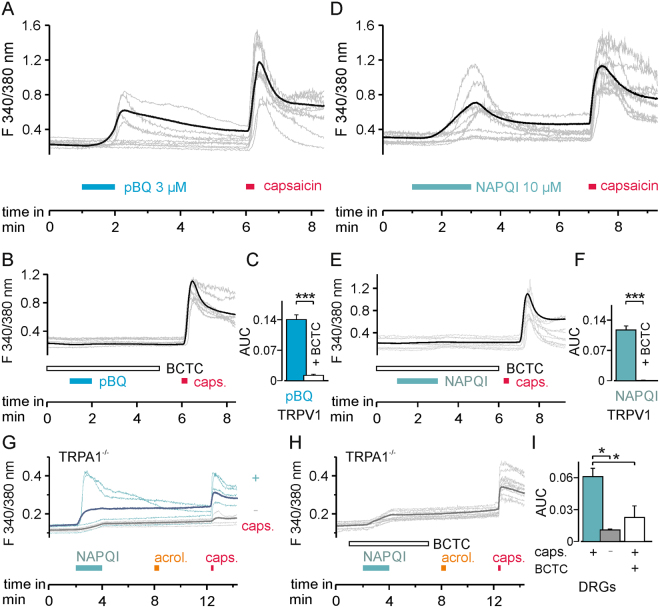
Figure 7.
pBQ and NAPQI induce a TRPV1-dependent rise in intracellular calcium. (A) In hTRPV1-expressing HEK 293 cells, 3 µM pBQ induced a rise in intracellular calcium in 36% of cells which also responded to capsaicin (n = 392). (B) Increase in intracellular calcium by pBQ was inhibited by the TRPV1 blocker BCTC (100 nM; n = 340). Responses are shown as mean of all cells responsive to pBQ (A) or capsaicin (B bold black trace), and calcium measurements of representative cells (thin grey traces). (C) Areas under the curve (AUC) of evoked calcium signals of all cells responding to pBQ: pBQ responses (blue bar) were significantly reduced by the TRPV1 channel blocker BCTC (white bar p < 0.001). (D) Similar to pBQ, 10 µM NAPQI evoked increases in intracellular calcium in hTRPV1-expressing cells (59%, n = 282). (A) These responses were inhibited by BCTC in 95% of capsaicin-responsive cells (n = 602). Bold black trace (mean) and thin grey traces (representative cells) of calcium measurements. (F) AUC of evoked calcium signals of all cells measured responding to NAPQI: NAPQI responses (cyan bar) were significantly reduced by BCTC (white bar; p < 0.001). (G) 10 µM NAPQI induced an increase in intracellular calcium in 15% of DRG neurons from TRPA1-knockout mice. Calcium responses of capsaicin-responding (blue, n = 216) and non-responding (gray, n = 166) subgroups of DRG neurons (bold = mean, thin = representative measurements of each group). (H) Co-application of the TRPV1 blocker BCTC reduced NAPQI-evoked calcium responses in DRG neurons from TRPA1-knockout mice (bold = mean of capsaicin-sensitive neurons, thin grey traces display representative measurements). (I) Comparison of the AUCs of calcium increase of the traces presented in (G and H). Increases in intracellular calcium following application of NAPQI are significantly inhibited in cells unresponsive to capsaicin (gray bar) or by TRPV1 block by BCTC (white bar; p = 0.00002).

