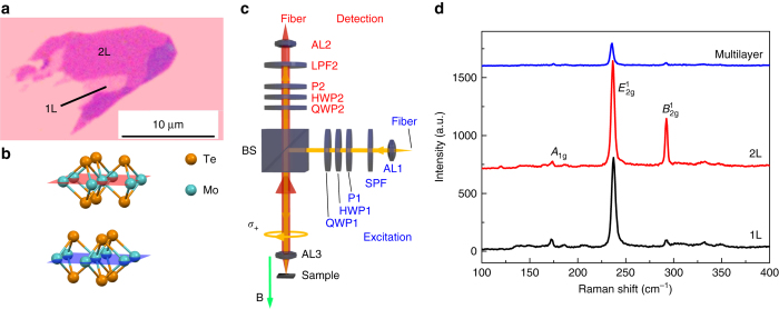
Fig. 1.
Sample characterization. a Optical microscope image of the MoTe2 monolayer and bilayer. b Crystal structure of a bilayer MoTe2. The two layers are rotated in-plane by 180° relative to each other. c Optical setup for the polarization-resolved PL spectroscopy. The optical components are: achromatic lenses (AL1-3), polarizers (P1 and P2), half-wave plates (HWP1 and HWP2), quarter-wave plates (QWP1 and QWP2), a short pass filter (SPF), a long pass filter (LPF), and a beam splitter (BS). The sample is placed in a helium bath cryostat with an out-of-plane magnetic field in a Faraday geometry. The green arrow shows a negative magnetic field. d Raman spectroscopy of the MoTe2 monolayer, bilayer, and multilayer. A 1g, , and represent different modes in Raman spectroscopy

