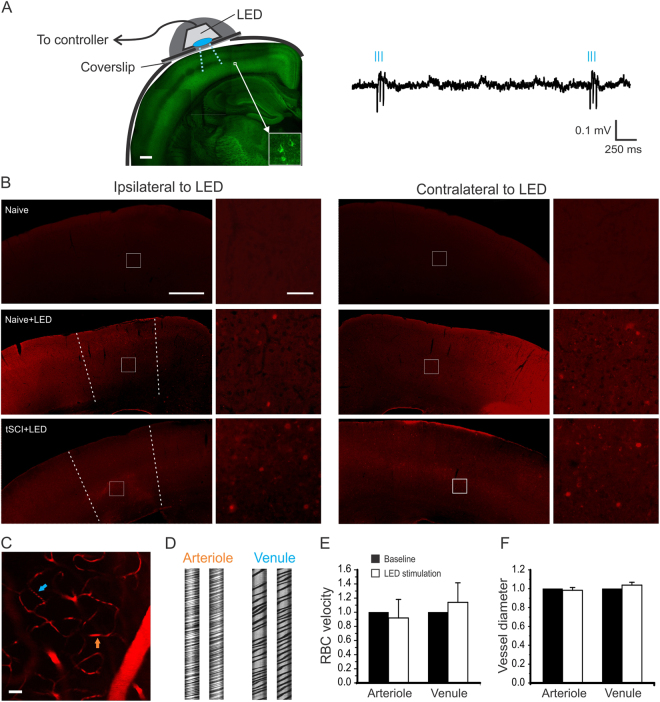
Figure 3.
Activation of S1 neurons using optogenetic stimulation in ChR2 mice. (A) Chronic optogenetic stimulation was made by implanting a blue LED (475 nm) on a cranial window over the S1 area of the Thy1-ChR2-YFP transgenic mice (left). About 1.5 mm diameter area of the window was exposed to the LED, with the surrounding area being blocked by opaque tape. Cortical expression of ChR2 is mostly in layer V pyramidal neurons (insert). The effectiveness of LED stimulation was confirmed by the evoked spikes of EEG recording (right) in vivo. (B) C-fos staining of cortical slices indicates activation of cortical neurons by the optogenetic stimulation. There was little staining in the cortex of naïve mice without optogenetic stimulation (top panel). For each row, c-fos staining of the ipsilateral (left two columns) and contralateral (right two columns) cortex are shown; enlarged views of the white square areas are shown to the right. The dotted lines indicate approximate borders of activated cortical neurons. After turning on blue light pulses (see methods) for 1.5 hours, there were c-fos positive cells in layer II/III and V in the S1 regions of both ipsilateral and contralateral hemispheres in uninjured ChR2 mice (middle panel). Similar bilateral c-fos staining pattern was seen in tSCI mice that received 1.5 hours LED stimulation (bottom panel). C–F No significant changes in cerebral vessel diameter and blood flow after light stimulation. Cerebral vessels were visualized by intravenous injection of sulforhodamine 101 in wild C57BL mice and imaged with two-photon microscopy (C). Cortical superficial arterioles and venules were identified and imaged in line scan mode (D). After 15 minutes of blue LED light stimulation, there were no significant changes in the velocities of red blood cells (RBC) and vessel diameters of arterioles and venules (E and F, n = 4 mice). Scale bars: A = 400 µm; B (1st and 3rd columns) = 500 µm; B (2nd and 4th columns) = 50 µm; C = 80 µm.

