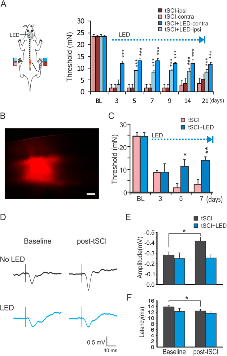
Figure 4.
Optogenetic stimulation of S1 prevents the development of mechanical hypersensitivity and reduces cortical hyperexcitability. (A) Optogenetic stimulation of the S1 in the tSCI model resulted in significant increases in withdrawal threshold forces of the bilateral hind-paws (***p < 0.001 when stimulated and unstimulated mice were compared; repeated measure ANOVA followed by Bonferroni test, n = 6 mice in each group). (B,C) Focal S1 optogenetic stimulation in virus transfected mice also induced analgesic effect. Following injection of ChR2-mCherry AAV into the S1 to infect layers II/III and V pyramidal neurons in a focal region (B), optogenetic stimulation with the same protocol also induced significant analgesic effect in the tSCI model (C). (*p < 0.05; **p < 0.01 when the tSCI + LED group was compared with the tSCI control group; Repeated measure ANOVA, n = 5 mice in each group). Scar bar: 300 µm. (D) Sample traces show that tSCI caused a high amplitude of cortical sensory-evoked potential, but optogenetic stimulation with LED for 5 days normalized it. (E,F) Group data showed that there were higher amplitudes of sensory-evoked response in tSCI group but not tSCI + LED group (E) and shorter latency in tSCI group but not tSCI + LED group (F). *p < 0.05, paired t-test in both E and F (tSCI group: n = 8 mice; tSCI + LED group: n = 5 mice).

