Figure 6.
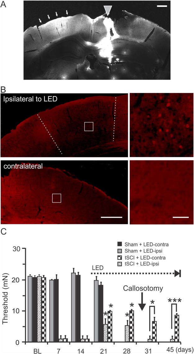
Analgesic effect of S1 optogenetic stimulation is mediated by cortical excitatory drive. (A). A corpus callosotomy was made using a “L-shaped” needle coated with a lipophilic fluorescent dye DiI, which labeled both local cut path (gray triangle) and truncated inter-hemispheric axons (arrows) of the intact cortical hemisphere (n = 3 mice). (B). In a ChR2 mouse that received callosotomy, c-fos staining shows that optogenetic stimulation for 1.5 hours activated only neurons ipsilateral to the LED (top images), but not neurons contralateral to it (bottom images) (n = 3 mice). The white squares indicate the location of the enlarged areas on the right. (C). Delayed optogenetic stimulation started on day 15 post-tSCI (dotted line) resulted in significant increases in withdrawal threshold forces of bilateral paws at days 21 and 28 (n = 4–5 mice in each group, p < 0.05). However, corpus callosotomy on day 28 (black arrow) resulted in a loss of analgesic effect of the ipsilateral hind-paw, but not the contralateral hind-paw, to optogenetic stimulation (p < 0.05-0.001 when compared between ipsilateral and contralateral paws, repeated- measure ANOVA). Scale bars in A and B (left column): 500 µm; B (right column): 50 µm.

