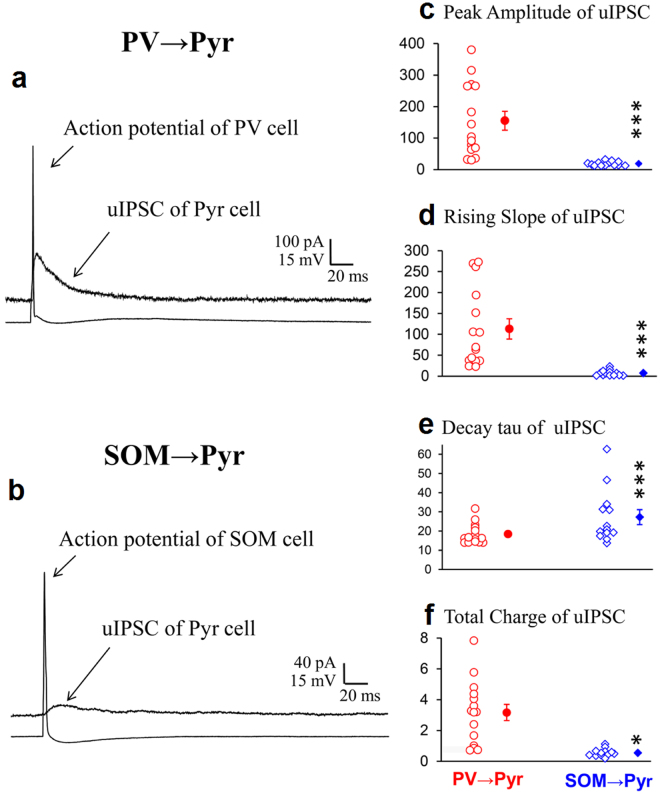
Figure 4.
uIPSCs of Pyr cells evoked by action potentials of PV or SOM neurons in vitro. (a,b) uIPSCs superimposed with action potential of a presynaptic PV and SOM neurons, respectively, recorded from Pyr cells in the slice preparation. Other conventions are the same as in Fig. 1a and b. (c–f) Peak amplitude, rising slope, decay tau and total charge of uIPSCs evoked by action potentials of PV neurons (left) and those evoked by action potentials of SOM neurons (right). Triple and single asterisks indicate that the difference in the mean between the left and right columns is statistically significant at p < 0.001 and p < 0.05, respectively (unpaired t-test).

